Kinematics and kinetics of motion
Understanding the physiological locomotion and gait of dogs is essential for the diagnosis of many musculoskeletal and neurological diseases. Gait assessment should be performed prior to any orthopaedic, neurological or physiotherapy examination.
The gait assessment usually involves visual, i.e. subjective observation of the dog from several angles while walking and trotting on a flat surface. Lameness can often already be detected during gait assessment with the trained eye. However, more subtle lameness may not be apparent during subjective gait assessment and may be difficult to detect. Recently, new proven technologies for objective gait analysis have become available to help veterinarians and physiotherapists quantify gait characteristics, which can greatly assist in detecting subtle lameness and response to various treatments (Carr B.J., 2016).
It is essential to have the means for objective gait analysis, as subjective gait assessment is difficult and unreliable. Objective analysis is particularly important in developing treatment plans and monitoring patient progress. Many methods of gait analysis have been developed (Table 1).
Table 1: Methods of gait analysis of dogs
Subjective |
Visual observation of gait (e.g. numerical rating scale, visual analogue scale) |
Objective |
Kinematic analysis of gait |
Kinetic gait analysis (force plate analysis) |
Spatiotemporal gait analysis (pressure sensing walkways) |
Kinematics
Kinematic gait analysis quantifies the positions, velocities, accelerations/decelerations and angles of various anatomical structures in space. Most kinematic gait analysis systems use coloured, retroreflective or light emitting diode (LED) markers to identify specific anatomical landmarks.
The most common sites where markers are placed include the dorsal margin of the scapula, the acromion/greater tubercle, the lateral epicondyle of the humerus, the ulnar styloid process, the iliac crest, the greater trochanter of the femur, the femorotibial joint, the lateral malleolus of the distal tibia, and the spinal process at the T13 vertebra.
Usually, the markers are attached after the skin has been shaved and cleaned with alcohol; then the marker and adhesive are pressed directly onto the skin. If necessary, the marker can be further secured with tape.
As the dog walks, a number of cameras track the movement of the markers. The location of the markers over time is then used to create a two- or three-dimensional model of the dog's gait, with calculations of bone and joint fluctuations. Kinematic parameters include displacements, angular velocities and range of motion:
- Shift is the distance recorded when the marker changes position.
- Angular velocity is the speed at which this change occurs.
- Range of motion is calculated from the displacement at the specific joint.
Although a wealth of information can be gained from this form of gait analysis, one of the main limitations is the variation in anatomical structures between and within breeds. Other limitations include the possibility of skin movement and the accuracy and repeatability of precise marker placement.
Kinetics
Kinetic gait analysis measures the ground reaction forces that result from an individual's stride. The most commonly used method of kinetic gait analysis is force plate analysis, in which metal plates are attached to the floor or pavement to measure ground reaction forces. The forces are measured in three dimensions: vertical, craniocaudal and mediolateral.
Table 2: Types of Ground Reaction Forces (GRF)
- Peak vertical force - PVF - Vertical impulse - Vertical impulse - VI - Rising and falling slope - Braking force - Braking force - Braking impulse - Propulsive force - Propulsive force - Driving impulse - Propulsive impulse - Mediolateral force - Mediolateral force |
These forces are often presented graphically, with the peak forces being the maximum forces generated in the gait phase described, represented by a force-time curve. The impulse is then represented as the area under the force-time curve.
- The peak vertical force (PVF) is the single largest force during the standing phase and represents only one data point on the force-time curve.
- The vertical impulse (VI) can be derived by calculating the area under the vertical force curve using time.
- PVF and VI are the two most commonly used indicators for lameness detection, and in general, a dog with lameness has a lower PVF and VI on a given limb
While braking, propulsive, and mediolateral forces may be useful in assessing locomotion mechanisms, they are not commonly used for diagnostic purposes or outcome assessment. Force plate measurements are still the most widely used and validated quantitative application of gait in veterinary medicine. Therefore, force plate analysis is considered the optimal approach to quantify gait characteristics using objective gait analysis. However, force plate analysis also has its disadvantages. Limitations include:
- Inability to measure stride or step length
- Need for consistent speed, long dedicated pavement and multiple attempts
- Difficult to assemble, disassemble and move
- Software complexity and data analysis
- Cost and impracticality for clinical practice
Spatiotemporal analysis of gait using pressure sensing carpets
Pressure sensing carpets have been validated for the analysis of normal and abnormal gait in dogs. This information helps in the diagnosis of orthopedic, muscular and neurological disorders that affect gait. These measurements provide new information on the temporal and spatial characteristics of gait.
Table 3: Description of the measurements calculated in the spatio-temporal analysis of gait
deadline | Definitions |
Standing time - Stance time | Stance phase in the gait cycle and paw to ground contact time |
Swing time - Swing time | The swing phase of the gait cycle and the time the paw is in the air. |
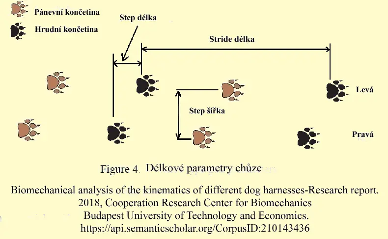
Stride length - Stride length | Distance from one footstep to the next footstep of the same limb |
Step length - Step lenght | Distance between the caudal point of one foot and the caudal point of the opposite foot |
Total pressure index - Total pressure index | The sum of the maximum pressure values recorded from each activated sensor by the paw in contact with the pad; related but not equal to the maximum vertical force. |
The portable pressure walkway system was originally developed for use in human medicine and has subsequently been modified and validated for use in veterinary medicine. Previous studies have established a protocol for spatiotemporal analysis and determined reference values and symmetry ratios for different breeds. Spatiotemporal gait analysis uses a pressure-sensing pad and a computer software system to calculate speed, standing time, swing time, stride length, stride length, and total pressure index.
The forces applied to change speed, change direction or maintain balance can interfere with and complicate the interpretation of the measurement. A pressure sensing walkway does not measure the force directly, but measures the effect of these forces. Therefore, by limiting excessive external influences, measurements can be more representative of the dog's actual gait.
As with other gait analysis systems, there are advantages and disadvantages to using a walkway with a pressure sensor. The advantages include:
- No size restrictions
- Multiple measurements from one pass
- Determination of stride length and step length
- Information about the location of the limbs
- User-friendly software
- Portability.
However, as with any other system, there are disadvantages, including:
- Ability to measure only the total ground reaction force
- Inability to separate 3 dimensions as in force plate analysis.
- Cost.
As more and more dogs participate in activities with their owners, dog sports or have different work roles, it is essential for owners and veterinarians to understand dog gait. Early signs of lameness can be as subtle as a shortened stride or a shorter standing time on the injured leg. Subjective and objective gait analysis is important not only for diagnosis but also for monitoring the progress of treatment.
Carr B.J., Dycus D.L. Canine Gait Analysis. Today´s Veterinary Practice, www.tvpjournal. com, March/April 2016, 93 - 100

