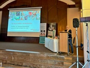
Search
Close this search box.
a2170-01-logo-box-200px
AK Vet Med

Articles

Physiotherapy

Retrogen FGF4 and chondrodystrophy

Retrogen FGF4 on canine chromosome CFA12 is responsible for chondrodystrophy and intervertebral disc disease in dogs.

Chondrodystrophy in dogs is defined by dysplastic, shortened long bones and premature degeneration and calcification of the intervertebral discs. It is a common phenotype in many of the most popular dog breeds, including the Dachshund, Pekingese, Beagle, and French Bulldog.
Histopathological analysis of the bones of puppies of these breeds showed that their short stature is caused by a defect in endochondral ossification, a process in which the cartilage of the bones in the developing limb is replaced. The growth plates of long bones show disorganisation of the proliferation zone and a reduction in the depth of the maturation zone. In addition to long bones, there are similar but more subtle changes in the endochondral ossification of vertebral bodies. Recent studies have shown that in advanced stages of degeneration in nonchondrodystrophic dogs, notochordal cells are also replaced by chondrocyte-like cells, similar to those in chondrodystrophic dogs, although this does not occur until older age.
The intervertebral disc (IVD), located between the vertebral bodies, consists of an outer fibrous ring, the annulus fibrosus, made up of 70 % collagen, and an inner gel pad, which is a remnant of the embryonic notochord, the nucleus pulposus. Together, these structures and the cartilaginous vertebral endplates allow the spine to be flexible. In chondrodystrophic individuals, the nucleus pulposus is gradually replaced by chondrocyte-like cells as part of chondroid metaplasia (or metamorphosis), which occurs between birth and 1 year of age.
Hansen described two different types of intervertebral disc prolapse in dogs as type I and type II. Type I occurs exclusively in chondrodystrophic breeds and is characterized by premature degeneration of all discs in young dogs. In contrast, type II occurs in older dogs and is usually limited to a single disc with partial protrusion. In type I disc disease, the calcified nucleus may erupt explosively through the anulus into the spinal canal, leading to inflammation and hemorrhage and causing severe pain and neurologic dysfunction. In population studies in veterinary hospitals, breeds found to have a significantly increased risk of intervertebral disc disease include the beagle, cocker spaniel, dachshund, French bulldog, lhasa apso, Pekinese, Pembroke Welsh corgi and shih tzu. Pet insurance data suggest a conservative "lifetime prevalence" of intervertebral disc disease in dogs of 3.5 % in the general population, however, the chondrodystrophic breeds at highest risk, the Dachshund has a lifetime prevalence of this disease of between 20-62 % with a 24% mortality rate.
In 2009, Parker et al. investigated the genetic basis of extreme differences in limb length in dogs using the method of association across breeds. They found that a retrogenic FGF4 insertion on canine chromosome CFA18 from the parental copy of the FGF4 locus is responsible for the "chondrodysplasia" phenotype in a number of breeds such as the Basset Hound, Pembroke Welsh Corgi and Dachshund. However, insertion of the FGF4 retrogene on canine chromosome CFA18 failed to explain breeds such as the American Cocker Spaniel, Beagle and French Bulldog, which, in addition to the Dachshund, were originally classified as chondrodystrophic based on histopathological and morphological analysis by Hansen and Braund. The FGF gene family has similarly been implicated in skeletal dysplasia in humans, with mutations in the FGFR3 gene found to be responsible for achondrodysplasia, the most common form of dwarfism, characterized by shortened limbs and abnormal vertebrae. FGF genes are involved in a number of embryological developmental processes and specific levels of ligand and receptor are crucial for proper growth and development.
In this study, based on whole-genome association analysis in a cohort of Nova Scotia duck tolling retrievers (NSDTR) with and without severe skeletal dysplasia, a significant association was found on the canine chromosome CFA12, where 1.9 Mbases were found to be common to chondrodystrophic breeds. Subsequent genome-wide association analysis of Hansen type I intervertebral disc disease across breeds localized the same 1.9 Mb region on canine chromosome CFA12, suggesting that the locus responsible for skeletal dysplasia in NSDTR is also responsible for type I intervertebral disc disease and the chondrodystrophic phenotype across dog breeds. Previous genetic investigations of intervertebral disc disease in Dachshunds and limb length morphology in Portuguese Water Dogs identified the same locus on the canine chromosome CFA12; however, neither study reported a causative mutation. Here, we used paired-end genome sequencing to reveal a second retrogenic FGF4 insertion in the canine genome and showed that it is responsible not only for skeletal dysplasia in NSDTR but also for chondrodystrophy, including a predisposition to Hansen's type I intervertebral disc disease, in all dog breeds.
Independent genome-wide association analyses for skeletal (short limb) dysplasia within a breed and intervertebral disc disease across breeds identified a significant association with the same region on canine chromosome CFA12. Whole genome sequencing identified a highly expressed FGF4 retrogene in this common region. The FGF4 retrogene segregated with limb length and had an odds ratio of 51.23 (95% CI =46.69, 56.20) for intervertebral disc disease. (Translator's note: genetic segregation involves the distribution of genes from parents to children). Long bone length in dogs is a unique example of a disease with multiple retrocopies of the same parental gene causing the disease. FGF abnormalities have been associated with skeletal dysplasia in humans, and our findings present opportunities for selective elimination of a medically and financially significant disease in dogs, as well as further understanding of the increasing complexity of retrogene biology.

Brown EA, Dickinson PJ, Mansour T, Sturges BK, Aguilar M, Young AE, Korff C, Lind J, Ettinger CL, Varon S, Pollard R, Brown CT, Raudsepp T, Bannasch DL. FGF4 retrogene on CFA12 is responsible for chondrodystrophy and intervertebral disc disease in dogs. Proc Natl Acad Sci U S A. 2017 Oct 24;114(43):11476-11481. doi: 10.1073/pnas.1709082114. Epub 2017 Oct 11. PMID: 29073074; PMCID: PMC5664524.

Continue reading "
Physiotherapy

Agility - injuries

   Little is known about the risk of injury in dogs participating in agility competitions. The aim of this study was to identify factors that increase the risk of injury in dogs

Continue reading "
Events

Congress Dogs in motion

   The main part of the physiotherapist's work is the musculoskeletal system and everything related to it. But can we look at it correctly? We've been taught at various

Continue reading "
Physiotherapy

Rupture of the cranial cruciate ligament

   Rupture of the cranial cruciate ligament (CrCL) is one of the most common orthopaedic diseases in veterinary medicine. The anterior cruciate ligament plays a critical role in the stability and biomechanics of the

Continue reading "
Physiotherapy

Osteoarthritis

   Osteoarthritis is a common and debilitating disease of domestic dogs. In addition to pharmaceutical interventions and weight reduction, exercise and physiotherapy are important measures to maintain mobility and

Continue reading "
Physiotherapy

Injury of the plexus brachialis

SUMMARY This article describes the association with plexus brachialis injury in dogs, especially with regard to prognosis. The final prognosis should be pronounced up to 6 weeks after

Continue reading "